3D/4D Ultrasound

3dultrasound
Contact us ANH 3D

What to do when and why ?

Ultrasound

Ideal schedule of pregnancy scans

Monitoring your pregnancy with timely scans ensures both your health and your baby’s well-being. Each scan is designed to check the healthy...
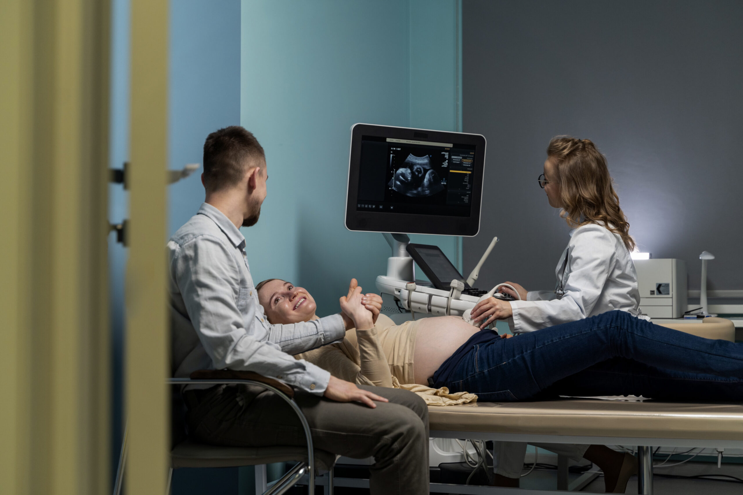
Ideal schedule of pregnancy scans

Monitoring your pregnancy with timely scans ensures both your health and your baby’s well-being. Each scan is designed to check the healthy development of your baby, detect any early concerns, and guide you through a safe pregnancy journey. You should do the following scans on time.

Ultrasound

Dating scan – 6 – 8 weeks

This is the first scan of pregnancy.The aim of this scan is to establish

  1. Location of the Gestation.

That is to look for...

Dating Scan

This is the first scan of pregnancy. The aim of this scan is to establish:

  1. Location of the Gestation Sac: Determines whether it is intrauterine or extrauterine. This is to look for ectopic pregnancy, which can potentially be life-threatening. Ideally, the gestation sac is implanted within the uterine cavity. Sometimes, the pregnancy gets implanted in the fallopian tubes, which is potentially life-threatening for the mother.
  2. Viability of the Foetus: This basically means that we are looking for the heartbeat or cardiac activity of the newly implanted baby.
  3. Dating the Pregnancy: Before ultrasounds, the only way to know pregnancy timing was by LMP (last menstrual period). Now ultrasounds can accurately determine the age of the foetus. This scan checks the growth of the baby to see if conception aligns with the LMP. Even if ultrasound dates do not match LMP dates, the scan can reassign the expected delivery date, helping in future scans to monitor growth and detect any issues.

The blood tests usually prescribed along with this scan are generally routine checkup tests.

Ultrasound

Level I Scan - NT/NB scan – 11- 14 weeks

NT stands for nuchal translucency. NB stands for nasal bone.Somehow for a layman seems pretty weird looking specifically for the nasal bone...

NT/NB Scan

NT stands for Nuchal Translucency. NB stands for Nasal Bone.

For a layperson, it may seem unusual to specifically check for the nasal bone or nuchal translucency. But for your gynaecologist, this is one of the most important tests for the future of the pregnancy. It is also known as the Level I Anomaly Scan. With a trained eye, it can provide the first glimpse into any potential problem with the child. It is the first hurdle the pregnancy needs to pass after the dating scan to look for ectopic pregnancy.

The NT scan properly measures the nuchal translucency, which is a small thickness behind the baby’s neck, and also examines the nasal bone. These are markers for common genetic syndromes, most notably Down’s syndrome. It is a statistical analysis — increased NT thickness or absent nasal bone may indicate a higher risk of genetic anomalies.

Genetic problems mostly cannot be solved with medicines or surgeries, as they are inherent flaws in the genetic makeup.

When to Do NT Scan?

The ideal time for this scan is when the length of the baby is between 45-85 mm, which corresponds to 11-14 weeks of pregnancy. Our radiologists prefer performing it around 12-13 weeks.

What is Dual Marker?

Dual marker is a blood test that examines hormonal markers in the maternal blood. When combined with the NT scan, the values of these hormones become a potent and fairly accurate statistical tool to detect genetic anomalies.

Our radiologists are Fetal Medicine Foundation (FMF), UK certified, and they ensure perfection, capturing the ideal section where the nasal bone, nuchal translucency, and intracranial translucency are clearly visible.

“Watching Dr Priyanka do NT scan is both learning and trust that we are in the safest of hands.” — Dr Nivetha N, Radiologist and patient

Ultrasound

Level 2 anomaly scan - TIFFA- CMF - 18 – 22 weeks

The TIFFA (Targeted Imaging for Fetal Anomalies) Scan, also known as the Level 2 Anomaly Scan, is a detailed ultrasound...

TIFFA (Targeted Imaging for Fetal Anomalies) Scan

The TIFFA (Targeted Imaging for Fetal Anomalies) Scan, also known as the Level 2 Anomaly Scan, is a detailed ultrasound performed between 18 and 22 weeks of pregnancy. It provides a complete assessment of your baby’s development and checks for any structural or congenital abnormalities. The CMF (Cranio-Maxillofacial) component specifically measures the face and skull and helps in detecting conditions such as cleft lip, palate, or other craniofacial anomalies.

Importance of Level 2 Anomaly Scan

  • Detailed Fetal Anatomy Check: It examines the vital organs of your baby, such as the brain, heart, kidneys, spine, limbs, and face.
  • Early Detection of Anomalies: Identify the congenital abnormalities and allow for timely medical advice.
  • Placenta & Amniotic Fluid Assessment: It ensures the placenta is functioning well and amniotic fluid levels are normal.
  • Gender Determination: Often provides an opportunity to know the baby’s gender (optional).

When is it recommended?

This scan is recommended between 18 and 22 weeks of pregnancy because this is the optimal time for accurate visualization of fetal structures. It is safe and uses advanced ultrasound technology to provide high-resolution images.

Ultrasound

Fetal ECHO – 22- 24 weeks

A Fetal Echocardiography, which is mostly known as Fetal Echo, is an ultrasound performed between 22 and 24 weeks of pregnancy...

Fetal ECHO – 22-24 weeks

A Fetal Echocardiography, which is mostly known as Fetal Echo, is an ultrasound performed between 22 and 24 weeks of pregnancy to determine your baby’s heart in depth. This is not a routine scan, as it focuses only on the fetal heart’s structure and function, ensuring it is developing normally.

Importance of Fetal ECHO Test

  • Detects Congenital Heart Defects (CHD): This test helps in identifying the heart abnormalities early, which are the most common birth defects.
  • Detailed Heart Examination: It assesses the chambers, valves, blood flow, and rhythm of the fetal heart.
  • Early Medical Planning: During this test, if any abnormality is detected, your doctor can plan treatment or intervention after birth.
  • Safe & Non-Invasive: It uses ultrasound waves with no radiation exposure, which makes it safe for both mother and baby.

Who needs a Fetal ECHO?

It is useful for all pregnancies, but it is highly recommended if:

  • There is a family history of congenital heart disease.
  • The mother has diabetes, lupus, or other medical conditions.
  • An abnormality is suspected in the routine anomaly scan.
  • The pregnancy involves IVF or multiple fetuses (twins/triplets).
Ultrasound

Interval Growth Scan

An interval growth scan is an ultrasound performed after 28 weeks of pregnancy, or in the second or third trimester. It is performed...

Interval Growth Scan

An interval growth scan is an ultrasound performed after 28 weeks of pregnancy, or in the second or third trimester. It is performed to monitor your baby’s growth and development over time. Unlike other routine scans, this test focuses on tracking the baby’s size, weight, and overall well-being at regular intervals.

It is important because it:

  • Monitors Baby’s Growth: This test checks that the baby is growing according to the expected pattern.
  • Assesses Amniotic Fluid & Placenta: It evaluates the placenta’s health and measures amniotic fluid levels.
  • Detects Growth Restrictions (IUGR): Identifies if the baby is smaller than expected or not receiving enough nutrition.
  • Checks Baby’s Position: It helps the doctors to know if the baby is in a cephalic (head-down) or breech position closer to delivery.
  • Supports High-Risk Pregnancies: This test is essential for mothers with diabetes, hypertension, or previous pregnancy complications.
Ultrasound

3D/4D/5D scan – 26 – 28 weeks

3D technology uses an added dimension to a usual 2D images for better visualisation and life like pictures of your baby...

3D/4D Ultrasound

3D technology adds an extra dimension to the usual 2D images for better visualisation and life-like pictures of your baby.

ANH is a pioneer in 3D/4D/5D ultrasound technology services, with the largest dedicated and trained fetal medicine specialists team.

The best time to do the 3D/4D scan is between 20-26 weeks. It generally coincides with the Level II Anomaly Scan and Fetal Echo.

Medically, it is used to look for cleft lip, cleft palate, binders facies, and a host of other structural anomalies. For parents, it provides the first glimpse into their upcoming baby.

What is the difference between 3D and 4D scan?

  • 3D Scan: Provides three dimensions — length, breadth, and height — offering life-like images as you would see with your eyes.
  • 4D Scan: Adds a fourth dimension — time — which creates a moving picture or video of your baby.
Ultrasound

Fetal Dopplers – 28- 36 weeks (as required)

Dopplers are a way of assessing the blood flow. This exam is crucial for timing the delivery.In fetal Dopplers...

Fetal Dopplers

Dopplers are a way of assessing the blood flow. This exam is crucial for timing the delivery.

In fetal Dopplers, we assess the blood flow going from the mother to the baby — namely from mother to placenta through the uterine arteries, and from the placenta to the baby via the umbilical arteries, Ductus venosus, aortic isthmus, and middle cerebral artery of the baby’s brain.

All these parameters give us an idea of when to deliver the baby and whether the baby is facing any imminent suffocation. They provide tools to predict any future derangement in the blood supply of the baby.

These assessments become especially important when one of the parameters is abnormal — that is the time when ANH radiologists come into play.

Patients in our antenatal care consistently show more than average fetal growth and weight gain due to our timely interventions.

At ANH, your Dopplers start happening from the second scan itself. During the NT/NB scan, ANH radiologists take into account the uterine arteries Doppler. During the Level II Anomaly Scan, uterine arterial Doppler and Ductus venosus are assessed.

Full fetal Dopplers are done post 28 weeks.

Ultrasound

Biophysical profile - After 28 weeks as required

A Biophysical Profile (BPP) is an advanced test that is performed after 28 weeks of pregnancy to evaluate the overall well-being of your baby...

Biophysical Profile - After 28 weeks as required

A Biophysical Profile (BPP) is an advanced test that is performed after 28 weeks of pregnancy to evaluate the overall well-being of your baby. It combines an ultrasound scan with a non-stress test (NST) to monitor both physical activity and oxygen supply.

This scan aims to establish:

  • Fetal Breathing Movements: Checks if the baby is practicing breathing inside the womb.
  • Fetal Body Movements & Muscle Tone: Observes the baby’s movements, stretching, and activity levels.
  • Amniotic Fluid Volume: Measures fluid levels, ensuring the baby has a safe environment for growth.
  • Heart Rate Monitoring: Evaluates the heart rate of the baby and checks the response to movements.
  • Overall Well-being: Detects signs of fetal distress and helps doctors decide if early delivery may be required.

Other available pregnancy scans

Ultrasound

Early level II anomaly scan – 16 weeks (in case of high risk on NT/NB scan + Dual marker)

This is without question the most important scan of the entire pregnancy. This is when the baby is best visualised...

Early level II anomaly scan – 16 weeks (in case of high risk on NT/NB scan + Dual marker)

This is without question the most important scan of the entire pregnancy. This is when the baby is best visualised. Under expert hands like our radiologists, who have a humongous checklist of parameters with the latest global protocols, they will look for everything structurally — from the corpus callosum in the brain to anorectal formation. We look for more than 50 parameters to ensure that the baby has no structural issues.

This scan combined with the quadruple marker is considered the final test of the pregnancy.

What to do if Level II Scan is Abnormal?

  • Level II scan may reveal many problems, some of which require no intervention and are not worrisome, while others may be medically significant.
  • It is best to consult our fetal medicine team, as every case is unique.
  • Usually, higher tests are recommended such as NIPT or amniocentesis.

Amniocentesis Procedure

Amniocentesis is when our skillful fetal medicine specialists take a sample of fluid from around the baby, known as amniotic fluid. It is done under aseptic precautions carefully without harming the baby. In expert hands, chances of complications are very low.

Why is Amniotic Fluid Important for Testing?

  • At around 16 weeks, the baby sheds skin cells into the amniotic fluid, allowing direct access to the baby's cells without touching the baby.
  • This fluid can then be tested for genetic anomalies.
  • Karyotyping and QF PCR
  • Low resolution micro-array
  • High resolution micro-array
  • Whole exome sequencing

We hope and pray that you never require these tests, but if you ever do, ANH is the most trusted name in fetal interventions.

Ultrasound

Chorionic villi sampling – 12- 14 weeks

Chorionic Villus Sampling (CVS) is a prenatal diagnostic test usually performed between 12 and 14 weeks of pregnancy. It usually involves collecting a small sample...

Chorionic Villi Sampling – 12-14 weeks

Chorionic Villus Sampling (CVS) is a prenatal diagnostic test usually performed between 12 and 14 weeks of pregnancy. It usually involves collecting a small sample of placental tissue (chorionic villi) to test for genetic or chromosomal conditions in the baby.

Why is CVS important?

  • Early Genetic Diagnosis: It detects chromosomal abnormalities such as Down’s syndrome, Edwards’ syndrome, Patau’s syndrome, and other genetic conditions.
  • Family History Screening: This test is recommended if there is a family history of genetic disorders.
  • Alternative to Amniocentesis: CVS can be done earlier (in the first trimester) compared to amniocentesis, giving the parents more time for decisions.
  • Helps in High-Risk Pregnancies: It is suggested when screening tests show a higher risk.

Procedure

  • A fine needle is inserted through the abdomen (or sometimes the cervix) under ultrasound guidance.
  • The collected placental cells are then analysed in a laboratory.

Although it carries a small risk of miscarriage, it is safe when performed by an expert.

Ultrasound

Amniocentesis (invasive) - 16 weeks

Amniocentesis is a prenatal test usually performed around 16 weeks of pregnancy.
It involves withdrawing a small amount of amniotic fluid from...

Amniocentesis (Invasive) - 16 weeks

Amniocentesis is a prenatal test usually performed around 16 weeks of pregnancy. It involves withdrawing a small amount of amniotic fluid from the sac surrounding the baby using a fine needle under ultrasound guidance. The fluid contains fetal cells and chemicals that provide vital information about the baby’s genetic health.

Why is Amniocentesis important?

  • Detects Chromosomal Abnormalities: It identifies Down’s syndrome, Edwards’ syndrome, and Patau’s syndrome.
  • Genetic Disorder Testing: It helps in detecting inherited conditions such as thalassemia, sickle cell disease, and cystic fibrosis.
  • Neural Tube Defect Detection: It can identify conditions like spina bifida.
  • Confirms Abnormal Screening Results: It is usually done after a high-risk result in NT scan, double marker, or triple marker tests.
  • Recommended for High-Risk Cases: Advised for women over 35 years of age, or when earlier screening indicates a higher risk of chromosomal or genetic abnormalities.

Procedure

  • A fine needle is inserted into the uterus under ultrasound guidance.
  • A small amount of amniotic fluid is withdrawn and sent to a lab for testing.

The procedure is safe when performed by an expert, but like all invasive tests, it carries a small risk of miscarriage.

Ultrasound

NIPT - after 12 weeks

Non-Invasive Prenatal Testing (NIPT) is a safe, advanced blood test that is performed after 12 weeks of pregnancy. It analyzes fetal DNA circulating in the mother’s blood to assess the risk...

NIPT - After 12 Weeks

Non-Invasive Prenatal Testing (NIPT) is a safe, advanced blood test that is performed after 12 weeks of pregnancy. It analyzes fetal DNA circulating in the mother’s blood to assess the risk of chromosomal abnormalities in the baby.

Why is NIPT important?

  • Early Detection of Chromosomal Abnormalities: Screens for Down’s syndrome (Trisomy 21), Edwards’ syndrome (Trisomy 18), and Patau’s syndrome (Trisomy 13).
  • High Accuracy: Provides highly reliable results for common chromosomal conditions.
  • Safe and Non-Invasive: Requires only a maternal blood sample, avoiding risks associated with invasive tests like CVS or amniocentesis.
  • Peace of Mind: Helps expectant parents make informed decisions early in pregnancy.

When is it Recommended?

  • Performed after 12 weeks of pregnancy.
  • Recommended for women aged 35+, those with a history of chromosomal abnormalities in prior pregnancies, or high-risk screening results.